Pulse (blow, push) is a jerky, periodic oscillation of the vascular wall.
Distinguish:
Central pulse: pulse of the aorta, subclavian and carotid arteries;
Peripheral pulse: pulse of the temporal arteries and arteries of the extremities;
Capillary (precapillary) pulse;
venous pulse.
The study of the pulse is of great clinical importance, as it allows you to obtain very valuable and objective information about the state of central and peripheral hemodynamics and the state of other organs and systems.
Pulse Properties
The properties of the pulse of the peripheral arteries depend on:
- frequency, speed and force of contraction of the left ventricle;
- magnitude of stroke volume;
- elasticity of the vascular wall;
- patency of the vessel (the value of the inner diameter);
- the value of peripheral vascular resistance.
The quality of the pulse should be evaluated strictly according to the following scheme:
- the same pulse on symmetrical arteries;
- frequency pulse waves per minute;
- rhythm;
- pulse voltage;
- filling the pulse;
- the value of the pulse;
- the shape of the pulse;
- condition of the vascular wall (vessel elasticity).
These 8 properties of the pulse must be known impeccably.
Pulse uniformity
In a healthy person, the pulse on the radial arteries is the same on both sides. The difference is possible only with an atypical location of the radial artery, in which case the vessel can be found in an atypical place - lateral or medial. If this fails, then pathology is assumed.
Pathological reasons for the absence of a pulse on one side or different pulse sizes on symmetrical vessels are as follows:
- anomaly in the development of the vessel,
- inflammatory or atherosclerotic vascular disease,
- compression of the vessel by a scar,
- a tumor
- lymph node.
Having found a difference in the properties of the pulse, it is necessary to establish the level of damage to the vessel by examining the radial artery at an accessible level, then the ulnar, brachial, subclavian arteries.
After making sure that the pulse is the same on both hands, further research is carried out on one of them.
Pulse rate
The pulse rate depends on the heart rate. It is better to count the pulse rate in the patient's sitting position after 5 minutes of rest in order to exclude the influence of physical and emotional stress (meeting with the doctor, walking).
The pulse is counted in 30 seconds, but better in 1 minute.
In a healthy person at the age of 18-60 years, the pulse rate ranges from 60-80 beats per minute, in women the pulse is 6-8 beats per minute more often than in men of the same age.
Asthenics the pulse is somewhat more frequent than in hypersthenics of the same age.
In old age in some patients, the pulse rate increases, in some it becomes less frequent.
For tall people the pulse is more frequent than in short people of the same sex and age.
Well trained people have a decrease in heart rate of less than 60 beats per minute.
Every person the pulse rate changes from the position of the body - in a horizontal position, the pulse slows down, when moving from a horizontal to a sitting position, it quickens by 4-6 beats, when standing up, it still quickens by 6-8 beats per minute. Newly adopted horizontal position slows down again.
All fluctuations in heart rate depend on from the predominance of the sympathetic or parasympathetic division of the autonomic nervous system.
- During sleep, the pulse especially slows down.
- Emotional, physical stress, eating, abuse of tea, coffee, tonic drinks leads to an increase in the tone of the sympathetic nervous system and an increase in heart rate.
- The phase of respiration also affects the pulse rate, on inspiration the frequency increases, on exhalation it decreases, which reflects the state of the autonomic nervous system - on inspiration the tone of the vagus decreases, on expiration it increases.
A pulse over 80 beats per minute is called fast. tachyphygmia, as a reflection of tachycardia, pulse less than 60 - rare, bradysphygmia as a reflection of bradycardia.
In practice, the terms tachyphygmia and bradysphygmia have not taken root; doctors, with the indicated deviations in the pulse rate, use the terms tachycardia and bradycardia.
Frequent heart rate
A frequent pulse that is not provoked by physical, emotional, nutritional and drug stress (atropine, adrenaline, mezaton, etc.) most often reflects trouble in the body.
Tachycardia can be of extracardiac and cardiac origin.
Almost all cases of fever are accompanied by an increase in heart rate, an increase in body temperature by 1 degree leads to an increase in heart rate by 8-10 beats per minute.
The increase in heart rate occurs when painful sensations, in most infectious and inflammatory diseases, with anemia, surgical diseases and surgical interventions with thyrotoxicosis.
Tachycardia in the form of seizures is called paroxysmal tachycardia, while the pulse rate reaches 140-200 beats per minute.
rare pulse
A rare pulse is noted with a significant increase in the tone of the vagus for extracardiac reasons - intracranial trauma, some diseases gastrointestinal tract, liver, decreased function thyroid gland(myxedema), cachexia, starvation, meningitis, shock, rapid ascent blood pressure, taking digitalis preparations, beta-blockers, etc.
For cardiac reasons, a rare pulse (bradycardia) is observed with weakness sinus node, blockade of the conducting system, narrowing of the mouth of the aorta.
The pulse rate, especially in cases of slowing down and arrhythmia, must be compared with the number of heartbeats counted in 1 minute during auscultation of the heart.
The difference between the number of heartbeats and the pulse is called pulse deficit.
Pulse Rhythm
In a healthy person, pulse waves follow at regular intervals, at regular intervals. Such a pulse is called rhythmic, regular, while the heart rate can be different - normal, rapid, slow.
A pulse with uneven intervals is called arrhythmic, irregular. In healthy adolescents and young people with labile autonomic regulation of blood circulation, respiratory sinus arrhythmia is noted. At the beginning of expiration, due to an increase in the tone of the vagus nerve, there is a temporary slowdown in the rate of heart contractions, a slowdown in the pulse rate. During inspiration, there is a weakening of the influence of the vagus and the heart rate increases slightly, the pulse quickens. When holding the breath, such respiratory arrhythmia disappears.
An arrhythmic pulse is most often caused by heart disease. It is most clearly detected in such heart rhythm disturbances as extrasystole and atrial fibrillation.
Extrasystole is a premature contraction of the heart. After a normal pulse wave, a premature small pulse wave slips under the fingers, sometimes it is so small that it is not even perceived. It is followed by a long pause, after which there will be a large pulse wave due to a large stroke volume. Then again there is an alternation of normal pulse waves.
Extrasystoles can be repeated after 1 normal beat (bigeminia), after 2 trigeminia), etc.
Another common variant of an arrhythmic pulse is atrial fibrillation. It appears with a chaotic contraction of the heart ("nonsense of the heart").
Pulse waves on the vessels have an irregular, chaotic alternation, they are also different in size due to the different stroke volume.
The frequency of pulse waves can range from 50 to 160 per minute. If atrial fibrillation begins suddenly, then they talk about its paroxysm.
An arrhythmic pulse is called in cases of its sudden increase in a person at rest, up to a frequency of 140-180 beats per minute, that is, with paroxysmal tachycardia. Such an attack can just as suddenly stop. Arrhythmic include the so-called alternating or intermittent pulse, in which there is a correct alternation of large and small pulse waves. This is typical for severe myocardial diseases, combinations hypertension with tachycardia.
An irregular pulse is also observed in other rhythm disturbances: parasystole, sick sinus syndrome, sinus node failure, atrioventricular dissociation.
Pulse voltage
This property reflects intravascular pressure and the state of the vascular wall, its tone and density.
There are no objective criteria for assessing pulse tension, the technique is being worked out empirically in the study of healthy and sick people.
The degree of pulse tension is determined by the resistance of the vessel to the pressure of the finger.
When determining tension, the third, proximal finger (the one closest to the heart) gradually presses on the artery until the distally located fingers no longer feel the pulsation.
In a healthy person with a normal pulse tension, a moderate effort is required to clamp the vessel. The pulse of a healthy person is estimated as a pulse of satisfactory tension.
If significant strengthening is required and the vascular wall has a significant resistance to clamping, then they speak of a tense, hard pulse, which is typical for hypertension of any genesis, severe sclerosis, or vasospasm.
A decrease in vessel tension, slight squeezing of the pulse indicates a soft pulse, which is observed with a decrease in blood pressure, a decrease in vascular tone.
Filling the pulse
It is estimated by the magnitude of the fluctuation of the vascular wall in systole and diastole, that is, by the difference between the maximum and minimum volumes of the artery. Filling mainly depends on the magnitude of the stroke volume and the total mass of blood, its distribution.
The degree of filling of the pulse can be judged using the following technique.
The proximal finger pinches the vessel completely, the distally located fingers feel the empty vessel, determining the state of the vascular wall. Then the pressure of the proximal finger stops, and the distal fingers feel the amount of filling of the artery. Fluctuations in the filling of the vessel from zero to the maximum reflects the filling of the vessel.
Another method for assessing the filling of the pulse is based on determining the magnitude of the fluctuation of the vascular wall from the level of diastolic filling to the level of systolic. All fingers placed on the vessel do not exert pressure on it, but only lightly touch the surface of the vessel during diastole. In systole, at the time of the passage of the pulse wave, the fingers easily perceive the magnitude of the fluctuation of the vascular wall, that is, the filling of the vessel.
In a person with normal hemodynamics, the filling of the pulse is assessed as satisfactory. With emotional and physical stress, as well as for some time (3-5 minutes) after exercise, due to an increase in stroke volume, the pulse will be full.
A full pulse is noted in patients with a hyperkinetic type of blood circulation (NCD, hypertension), as well as in aortic insufficiency. Poor filling pulse - empty pulse - patients with severe hemodynamic disorders (collapse, shock, blood loss, myocardial insufficiency) have.
Pulse value
The value of the pulse is a reflection of the relationship of such properties of the pulse as filling and tension. It depends on the magnitude of the stroke volume, the tone of the vascular wall, its ability to elastic stretch in systole and fall in diastole, on the magnitude of blood pressure fluctuations in systole and diastole.
In a healthy person with satisfactory filling and tension of the pulse, the pulse value can be described as satisfactory. However, in practice, the magnitude of the pulse is spoken only when there are deviations in the form:
Large pulse (high pulse);
Small pulse (its extreme form is filiform).
big pulse occurs with increased stroke volume and reduced vascular tone. The fluctuation of the vascular wall under these conditions is significant, so a large pulse is also called high.
In healthy people, such a pulse can be felt after physical activity, baths, baths.
In pathology, patients with valve insufficiency, aorta, thyrotoxicosis, and fever have a large pulse. At arterial hypertension with a large difference between systolic and diastolic pressure (large pulse pressure), the pulse will also be large.
Small stroke volume of the left ventricle gives rise to a small amplitude of oscillation of the vascular wall in systole and diastole. An increase in vascular tone also leads to a decrease in the oscillation of the vascular wall during the cardiac cycle. All this fits into the concept of a small pulse, which patients have with such heart defects as narrowing of the aortic orifice, stenosis mitral valve. A small pulse is characteristic of acute cardiovascular vascular insufficiency.
In shock, acute heart and vascular insufficiency, massive blood loss, the pulse is so small that it is called a thready pulse.
Pulse shape
The shape of the pulse depends on the rate of pressure change in the arterial system during systole and diastole, which is reflected in the rate of rise and fall of the pulse wave.
The shape of the pulse also depends on the speed and duration of contraction of the left ventricle, the state of the vascular wall and its tone.
In a person with normal functioning of cardio-vascular system when evaluating the pulse, one usually does not talk about the shape of the pulse, although it could be called “normal”.
As options for the shape of the pulse, fast and slow pulses are distinguished.
In healthy people, only a fast pulse can be detected after physical and emotional stress. Fast and slow pulses are found in pathology.
Fast (short, jumping) pulse
Fast (short, jumping) pulse is characterized by a steep rise, a short plateau and a sharp decline in the pulse wave. Such a wave is usually high. A fast pulse is always detected with aortic valve insufficiency, in which there is an increased stroke volume, a large force and speed of contraction of the left ventricle in a short time, a large difference between systolic and diastolic pressure (diastolic may drop to zero).
A fast pulse occurs with reduced peripheral resistance (fever), with thyrotoxicosis, some forms of hypertension, nervous excitability, and anemia.
slow pulse
Slow pulse - the opposite of a fast one, characterized by a slow rise and fall of a low pulse wave, which is due to the slow rise and fall of blood pressure during the cardiac cycle. Such a pulse is due to a reduced rate of contraction and relaxation of the left ventricle, an increase in the duration of systole.
A slow pulse is observed when there is difficulty in expelling blood from the left ventricle due to an obstruction in the path of blood outflow into the aorta, which is characteristic of aortic stenosis, high diastolic hypertension. A slow pulse will also be small due to the limitation of the magnitude of the oscillation of the vascular wall.
Dicrotic pulse
A dicrotic pulse is one of the features of the pulse shape, when a short-term slight rise is felt on the falling part of the pulse wave, that is, the second wave, but of lesser height and strength.
An additional wave occurs when the tone of the peripheral arteries is weakened (fever, infectious diseases), it expresses the back wave of blood reflected by the closed aortic valves. This wave is the greater, the lower the tone of the arterial wall.
Dicrotic pulse reflects a decrease in peripheral vascular tone with preserved myocardial contractility.
The state of the vascular wall
The vascular wall is examined after complete clamping of the artery with a proximal finger, that is, an empty vessel is examined. Distally located fingers feel the wall by rolling through the vessel.
A normal vascular wall is either not palpable or is defined as a tender, soft, flattened band about 2–3 mm in diameter.
In old age, the vascular wall sclerotizes, becomes dense, palpable in the form of a cord, sometimes the vessel is convoluted, bumpy in the form of a rosary. A dense, poorly pulsating or non-pulsating artery occurs with Takayasu's disease (pulseless disease), which is caused by inflammation of the vascular wall, as well as vascular thrombosis.
Pulse deficit
Pulse deficit is a discrepancy between the number of heartbeats and the number of pulse waves.
This means that part of the pulse waves does not reach the periphery due to a sharply reduced stroke volume of individual heart contractions.
This happens with early extrasystoles and with atrial fibrillation.
Vibrations in the walls of blood vessels caused by the contraction of the heart. The arterial pulse is formed by fluctuations in blood pressure and blood supply in the arteries during the cardiac cycle. Normal heart rate is 60-80 beats per minute. Biology. Modern Encyclopedia
With every beat hearts a new portion of blood enters the arteries. If it were not for the elastic extensibility of the arterial system, this blood would flow through the peripheral vessels only during systole, and during diastole the blood flow would stop. The ability of the arteries to accommodate an additional volume of blood leads to a decrease in pulse fluctuations in blood flow until they completely disappear by the time the blood reaches the capillary bed. Thus, tissue blood flow is carried out continuously, with negligible pulse fluctuations.
The figure shows the recording of pulse blood pressure fluctuations at the beginning of the aorta. In healthy young people, the maximum, or systolic, pressure is approximately 120 mm Hg. Art., and the minimum, or diastolic, pressure is about 80 mm Hg. Art. The difference between systolic and diastolic pressure (about 40 mm Hg) is called pulse pressure. Two main factors influence the value of pulse pressure: (1) stroke volume of the heart; (2) compliance (extensibility) of the arterial system. A third, less important factor is the nature of the ejection of blood from the heart during systole.
In general, the more stroke volume, the more blood must fit into the arterial vessels during each heartbeat, therefore, the greater the systolic increase and diastolic decrease in pressure will be, which will lead to an increase in pulse pressure. Conversely, the lower the compliance of the arterial wall (i.e., the reserve capacity of the arterial system), the greater will be the rise in pressure with the same stroke volume of blood entering the arteries. In the middle part of the upper curve in the figure, it is shown that the pulse pressure in old age can increase by more than 2 times compared to the norm, because. arteries become stiffer as a result of atherosclerosis and their capacity is significantly reduced.
It can be said that pulse pressure is determined by the ratio of stroke volume to the reserve capacity of the arterial system. Any changes in hemodynamics that affect these two factors also affect the value of pulse pressure.
Changes in pulse pressure
Many hemodynamic disorders cause not only a change in the value of pulse pressure, but also a pattern of pulse pressure fluctuations. Particularly significant disorders are aortic stenosis, cleft ductus arteriosus, aortic valve insufficiency.
With aortic stenosis the diameter of the opening with open aortic valves is significantly reduced compared to the norm. The pulse pressure in the aorta also decreases, since there is a decrease in blood flow through the stenotic valves.
In case of non-closure of the arterial (Botallov) duct about half of the stroke volume of blood that should enter the aorta from the left ventricle immediately enters the pulmonary artery and pulmonary vascular system through the wide open duct. This is accompanied by a significant drop in diastolic pressure before each subsequent heartbeat.
With aortic valve insufficiency absent or incompletely closed aortic valves. Therefore, after each heartbeat, the blood that has entered the aorta immediately returns to the left ventricle. As a result, aortic pressure drops to zero during diastole. In addition, there is no incisura on the aortic pressure curve, because aortic valves do not close.
Since ancient times, the pulse on the hand has been measured in order to understand its effect on human health. They succeeded. In case of violation or change in blood circulation, blood loss, the “filling of the pulse” decreases.
Thanks to this knowledge, heart rate can now be easily controlled using the second hand of a watch.
In those ancient times, the study of the human pulse was a characteristic feature of medicine in various countries, but Chinese medicine approached it most specialized.
Further teachings of this direction developed everywhere in all countries, but to a large extent, Chinese medicine has retained the knowledge gained to this day, which makes it possible for modern science to investigate such facts in more detail.
Pulse - these are rhythmic or undulating oscillations of the walls of blood vessels, caused by the continuous work of the fibromuscular organ together with its contractions. Reasoning very clearly, this term means any change that is associated with the activity of the heart in the vascular system.
The fibromuscular organ often increases in size, especially when a person is engaged in training processes, and the heart also grows based on the growth of the person himself, taking into account physiology.
It is important to understand that the frequency of vascular oscillations, the rate, is relevant not only as an active regulator in controlling the activity of the heart, but also as an indicator of the level of physical fitness of the human body.
As practice shows, in a “normal” state, a person has readings that mean: the lower the pulsation, the better. Therefore, the "pulse is" arterial, venous, capillary, and the main way to study it is considered to be palpation - probing the arteries.
Arterial pulse and its features
It is customary to call the arterial pulse “such” tempo fluctuation that occurs in the walls of blood vessels, predetermined by the splash of blood from the fibromuscular organ into the arterial system and the variation in pressure in this system during the period of systole and diastole.
This pace is based on the perspective of large, medium-sized arteries that are unreasonably located, in a larger form responding to the active work of the fibromuscular organ. The constantly moving walls of the arteries form the flow of blood inside them, the pressure of which is accelerated by the rhythmic movement of the ventricles of the heart, i.e. its circulation.
The pulse wave passes through the vascular system unequally; as a result of the distribution of the blood flow, the pulse has the character of a slight delay with the time of the work (beat) of the heart. If you simultaneously look for a pulse on the carotid artery, then you need to take into account the work of the heart. The difference will not be noticeable, because the vessel is close, so there is a reaction to the release of blood.
Let's pay attention to the wrist, here is the radial artery, and the difference in the distribution of blood flow with the impacts of the fibromuscular organ is less than 1 second, so such an insignificant difference is not worth attention.
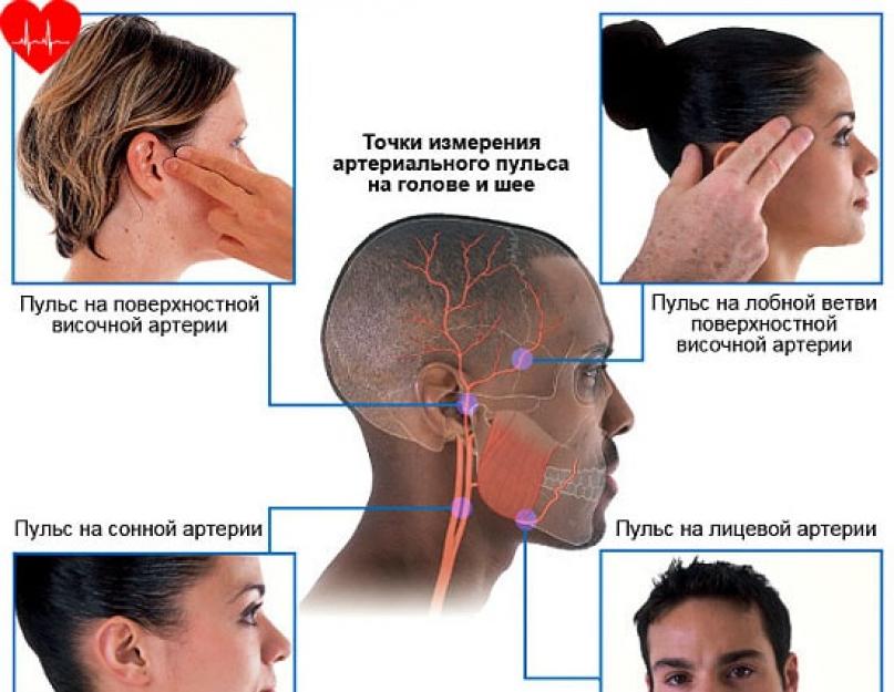
The most significant differences can be those actions when finding a pulsation on the foot is a fairly clear delay. From the specific vessels being measured, often the arterial is referred to as the peripheral or "central pulse". It is found on solid vessels - carotid, also called carotid arteries, in the aorta.
As many sources show, the pulse is found at the following points in the limbs:
- upper (arteries radial, axillary, brachial, as well as ulnar);
- lower (arteries of the foot, posterior tibial, popliteal and femoral);
- further head (superficial temporal, facial, carotid arteries).
Arterial compared to capillary and venous, more useful in diagnosis.
Exceptional Arterial Pulse Characteristics

There are such "characteristics of the arterial pulse" as:
- rhythm,
- filling,
- voltage,
- frequency,
- magnitude (height),
- speed (shape).
Let's note the rhythm as a value that is "determined" by time gaps (intervals) before and after the sequence of pulse waves.
Allocate arrhythmic and rhythmic. When pulse waves move alternately and mutually through identical time frames, the pulse is dated as rhythmic and vice versa, in another case, as arrhythmic.
Next, consider the filling of the arterial pulse - this is the amount (volume) of the available blood of the artery located at the height of the elevation of the pulsation on which the artery is located. They “distinguish” filiform (little perceptible), empty (poorly perceptible), full (overfilled), moderate.
“Pulse tension” is spoken of as the advantage of effort applied before the artery is unconditionally pressed. It happens soft, hard, moderate tension.
"Pulse rate" this is a value whose “parameter” is “determined” by the number of oscillations of the arterial walls in 1 time. It means how many beats per minute the fibromuscular organ does. It happens moderate (60-80 bpm), which means the value is "normal", rare (less than 60 bpm), frequent (more than 90 bpm).
There are some inversions: bradycardia (tightening of the heartbeat), tachycardia (rise of pulse waves).
Determining the frequency is of great practical importance in the registration of indications of clinics and physiology.
An interesting concept is the "pulse value" (height) - "defined" as the range of vibration of the edges of the vessels, this is the totality of the filling value along with its tension. The property, namely the "pulse value" is small, large, moderate.
The final concept is speed (form) - it means the haste of reforming the size of the vessel. Recognized by sphygmogram. The sphygmograph determines the increase and decrease of the emerging waves, after which it displays a “graph”, where there is a clear movement. Subdivided into fast, slow, dicrotic.
Capillary and venous pulses

Capillary and venous pulses are also important in diagnosis.
“The capillary pulse is” a predominantly undulating movement of the capillary walls. The real rate of movement of the walls of the capillaries is observed in the younger generation with fever and high climatic changes.
Enough it is manifested by a significant change in the color of the forehead, such a change occurs when it is affected by small mechanical movements.
It can also be observed on the surface of the face, especially the mucous membrane of the lips, with progressive pressure on a transparent glass object. The capillary rate becomes visible as a result of the heterogeneous degree of saturation of the veins (the cycle of systole and diastole) of the fibromuscular organ, which gives the right to the arterial knee of the capillaries to pulsate rapidly.
Patients suffering from significant diseases have a peculiar even interesting pulse, “which” is observed in the form of pupillary pulsation simultaneously with the heart rhythm.
The venous is "determined" by the action, the pace of which takes part in the veins that are not located near the stomachs of the heart, but are separated by a part of the capillary vessels. It is these veins that do not receive blood flow through shocks (blows), this is the reason for the absence of oscillations. On fair veins, sometimes, a rhythmic pulsation may occur. The jugular veins are influential in the manifestation of venous tempo.
During physical exercises and repeated psychological, emotional upheavals in people with a model (thin) body, this type of pulse shows itself in the picture under the guise of pulsating tourniquets, which is considered “normal”.

More than four thousand years ago, the ancient Egyptians knew the diagnosis of diseases by the pulse!
The arterial pulse is a rhythmic jerky oscillation of the vessel wall resulting from the ejection of blood from the heart into the arterial system. Pulse from lat. pulsus - push.
Physicians of antiquity paid great attention to the study of the properties of the pulse. The scientific basis of the doctrine of the pulse was received after the discovery of the circulatory system by Harvey. The invention of the sphygmograph and especially the introduction of modern methods of pulse recording (arteriopiezography, high-speed electrosphygmography, etc.) have significantly deepened knowledge in this area.
With each systole of the heart, a certain amount of blood is ejected into the aorta. This blood stretches the initial part of the elastic aorta and increases the pressure in it. This change in pressure propagates along the aorta and its branches to the arterioles. In the arterioles, the pulse wave stops, because. there is high muscle resistance. The propagation of the pulse wave occurs much faster than the blood flows. The pulse wave goes at a speed of 5-15 m / s, i.e. it runs 15 times faster than blood. That. the occurrence of a pulse is due to the fact that during the work of the heart, blood is pumped into the vessels inconsistently, but in portions. The study of the pulse allows you to judge the work of the left ventricle. The greater the systolic volume, the more elastic the artery, the greater the wall oscillations.
Vibrations of the walls of the arteries can be recorded using a sphygmograph. The recorded curve is called a sphygmogram. On the pulse recording curve - sphygmogram, an ascending knee is always visible - an anacrota, a plateau, a descending knee - a catacrot, a dicrotic rise and an incisura (notch).
Anacrota occurs due to an increase in pressure in the arteries and coincides in time with the phase of rapid expulsion of blood into the systole of the ventricles. At this time, the inflow of blood is greater than the outflow.
Plateau - coincides with the phase of slow expulsion of blood into the systole of the ventricles. At this time, the inflow of blood into the aorta is equal to the outflow. After systole, the semilunar valves close at the beginning of diastole. The inflow of blood stops, but the outflow continues. The outflow predominates, so the pressure gradually decreases. This causes catacrosis.
In the proto-diastolic interval (end of systole, beginning of diastole), when the pressure in the ventricles decreases, the blood tends back to the heart. Outflow is decreasing. An incisura occurs. During diastole of the ventricles, the semilunar valves close and, as a result of the impact on them, a new wave of blood outflow begins. There is a short-term wave of increased pressure in the aorta (dicrotic rise). After that, the catacrosis continues. The pressure in the aorta reaches the initial level. Outflow increases.
Pulse properties.
Most often, the pulse is examined on the radial artery (a.radialis). In this case, pay attention to the following properties of the pulse:
1. Pulse rate (HR). PE characterizes the heart rate. Normal PR = 60 - 80 beats / min. With an increase in HR over 90 beats / min, they speak of tachycardia. With a decrease (less than 60 beats / min) - about bradycardia.
Sometimes the left ventricle contracts so weakly that the pulse wave does not reach the periphery, then the number of pulse beats becomes less than the heart rate. This phenomenon is called bradysphygmia. The difference between heart rate and HR is called pulse deficit.
According to the state of emergency, you can judge what T a person has. An increase in T by 1 0 C leads to an increase in heart rate by 8 beats / min. The exception is the change in T in typhoid fever and peritonitis. With typhoid fever, there is a relative slowing of the pulse, with peritonitis - a relative increase.
2. The rhythm of the pulse. The pulse may be rhythmic or arrhythmic. If pulse beats follow one after another at regular intervals, then they speak of a correct, rhythmic pulse. If this period of time changes, then they speak of an incorrect pulse - the pulse is arrhythmic.
3. The speed of the pulse. The speed of the pulse is determined by the rate of rise and fall of pressure during the pulse wave. Depending on this indicator, a fast or slow pulse is distinguished.
A fast pulse is characterized by a rapid rise and a rapid decrease in pressure in the arteries. A fast pulse is observed with aortic valve insufficiency. A slow pulse is characterized by a slow rise and fall in pressure, i.e. when the arterial system slowly fills with blood. This happens with stenosis (narrowing) of the aortic valve, with weakness of the myocardium of the ventricle, fainting, collapse, etc.
4. Pulse tension. It is determined by the force that must be applied to completely stop the propagation of the pulse wave. Depending on this, a tense, hard pulse is distinguished, which is observed with hypertension, and an unstressed (soft) pulse, which occurs with hypotension.
5. Filling or amplitude of the pulse is a change in the diameter of the vessel during the pulse push. Depending on this indicator, a pulse with a large and small amplitude is distinguished, i.e. good and bad content. The filling of the pulse depends on the amount of blood ejected by the heart and on the elasticity of the vascular wall.
There are many more properties of the pulse, which you will get acquainted with in therapeutic departments.
Venous return.
One of the important indicators of systemic hemodynamics is the venous return of blood to the heart. It reflects the volume of venous blood flowing through the superior and inferior vena cava. Normally, the amount of blood flowing in 1 minute is equal to the IOC. The ratio of venous return and cardiac output is determined using special electromagnetic sensors.